

Oxytocin
Oxytocin
This batch of Oxytocin Peptide has been third party lab tested and verified for quality.
Contents: Oxytocin (Neuropeptide Hormone)
Form: Powder
Purity: 99.3%
View Third-Party Tests from Our Partners:
Couldn't load pickup availability
This product is Made, Tested & Shipped From United States.
Shipping & Delivery
We are committed to delivering fast, reliable, and transparent shipping for all orders. Please review our policy below for details on delivery times, tracking, and what to expect with every purchase.
View Full Shipping Policy

Verified+


You may also like
-
 SAVE 23%
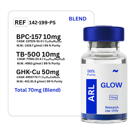
Glow BPC-157 + GHK-CU + TB-500
Regular price $139.00Regular price $139.00 Sale priceUnit price per$181.0023% -
 SAVE 23%
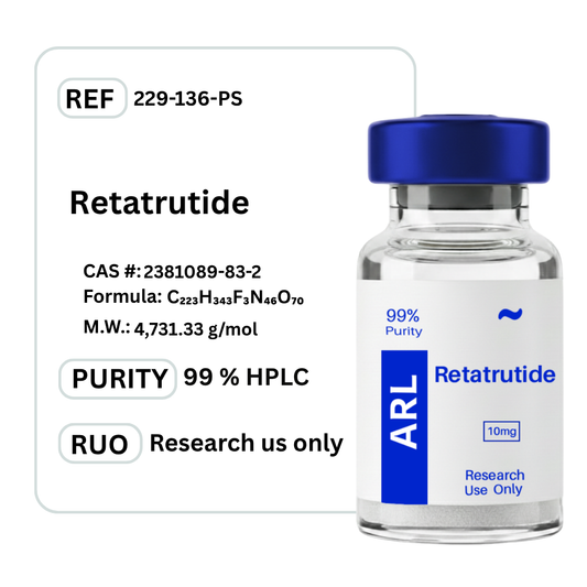
Retatrutide Triple Agonist
Regular price From $110.00Regular price From $110.00 Sale priceUnit price per$144.0023% -
 SAVE 7%
BPC-157 + TB-500
Regular price $118.00Regular price $118.00 Sale priceUnit price per$127.007% -
 SAVE 23%
Cagrilintide
Regular price $150.00Regular price $150.00 Sale priceUnit price per$197.0023% -
 SAVE 23%
CJC-1295 No DAC & Ipamorelin
Regular price $95.00Regular price $95.00 Sale priceUnit price per$124.0023% -
 SAVE 23%
Ipamorelin
Regular price $63.00Regular price $63.00 Sale priceUnit price per$82.0023% -
 SAVE 23%
Kisspeptin-10
Regular price $95.00Regular price $95.00 Sale priceUnit price per$124.0023% -
KLOW Blend - GHK-CU + TB-500 + BPC-157 + KPV 10mg
Regular price $200.00Regular price $200.00 Sale priceUnit price per$261.0023% -
 SAVE 22%
Reconstitution Solution
Regular price $27.00Regular price $27.00 Sale priceUnit price per$35.0022% -
 SAVE 23%
Semaglutide
Regular price From $42.00Regular price From $42.00 Sale priceUnit price per$55.0023% -
 SAVE 23%
Sermorelin
Regular price $70.00Regular price $70.00 Sale priceUnit price per$92.0023% -
 SAVE 23%
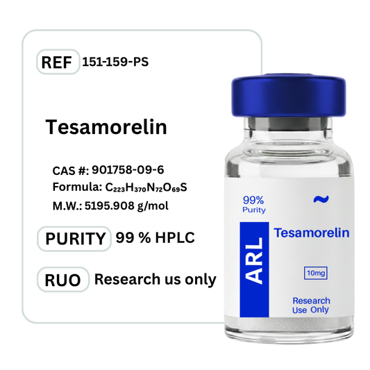
Tesamorelin
Regular price $110.00Regular price $110.00 Sale priceUnit price per$144.0023% -
 SAVE 23%
Tirzepatide
Regular price From $70.00Regular price From $70.00 Sale priceUnit price per$92.0023%


Frequently Asked Questions
Here you'll find answers to common questions.
Every vial we sell comes from a lab that follows current Good Manufacturing Practices (cGMP). That means each step of production is documented and controlled. Before a batch is released, it’s tested by independent third-party labs for purity, identity, and sterility. Certificates of analysis are available so you can see the exact test results.
Yes. The labs we work with use ISO-certified clean rooms where air quality, equipment, and handling procedures are tightly regulated. Staff are trained to pharmaceutical-grade standards. This ensures the peptides are produced in an environment that minimizes contamination risks.
Peptides in lyophilized (freeze-dried) form are stable at room temperature for transport. Once you receive them, refrigeration is recommended to maintain long-term integrity. We package every order securely to prevent damage and ship promptly, so your vials arrive in optimal condition.
We operate under strict in-house protocols that follow current Good Manufacturing Practices (cGMP). That means our team oversees the entire process from sourcing raw amino acids to the final lyophilized vial. Nothing is outsourced or repackaged. This gives us full control over purity, consistency, and sterility, and it’s why we can stand behind every single vial we ship.
Store them in the refrigerator, away from direct light and heat. If you need to keep them longer, some peptides can be stored frozen. Each vial comes with clear handling instructions so you know the proper conditions for stability.
The strongest proof is transparency. For every peptide, we can provide certificates of analysis, manufacturing documentation, and references to the published scientific research behind it. If you ever have questions, we’ll show you the data rather than ask you to take our word for it.
The difference is transparency. Most sites give you a product name and a price. We provide full batch testing, lab documentation, and direct access to certificates of analysis so you don’t have to guess what you’re getting. When you order from us, you know exactly what’s in the vial, where it was made, and how it was verified.